Follow me over to Substack!
Instead of making updates here, I’ve started a monthly newsletter to explore the role of art in science that will include recent work from time to time. Check it out if you’re interested, and if you like it, subscribe for free to receive one e-mail a month when I post a newsletter. https://maryoreilly.substack.com/
Thanks!
Encapsulated: A Love Letter to the Purdue Chemistry Department
I went to graduate school more or less because I wanted to be Jean Chmielewski. So when she asked me recently if I could create cover art for a themed issue of The Journal of Peptide Science, of course, I immediately agreed.
She and her graduate student Michael Jorgensen were publishing a paper in that issue on reversible encapsulation of cells in a 3D matrix that mimics their native environment — the extracellular matrix. Taking cells out of their native environment leaves them a bit lost, but Chmielewski and Jorgensen had engineered a support system for them.
I was a year and a half into my chemistry major at Purdue, a pre-med by default since I had no clue what chemists actually did, when a classmate told me that undergraduate research experience would look good on my medical school application. I asked an assistant professor named Bruce Morimoto if I could join his lab, and despite the fact that I naively told him my motivation for it, he welcomed me anyway. I spent the holidays reading his papers and then got to work.
The support system that Chmielewski and Jorgensen devised uses a helical peptide that gently twists together with two of its identical twins to form trimeric coiled coils like very short lengths of rope. They equipped each peptide with multiple metal-binding arms. When metal is added to the solution, each metal ion can bind to two separate arms, forming bridges between neighboring peptide trios that enable them to self-assemble into larger and larger 3D structures. When cells are present, these structures assemble themselves around the cells like a molecular scaffolding.
In the Morimoto Lab I was delivered into the capable hands of a warm and knowledgeable graduate student who taught me how to clone a gene and sequence DNA, decoding it old-school style from spots on a sheet of photographic film the size of a Denny’s menu. She invited me to her home and cooked Indian food for me — the first and still the best I’ve ever had. But about 6 months in, Morimoto left academia for biotech.
At any moment, the peptidic 3D support system can be immediately disassembled simply by adding a metal chelator that sponges up the bridging metal ions, dispersing the trimeric peptidic coiled coils and leaving the cells to fend for themselves again.
I met Professor Chmielewski at a summer BBQ where in between volleyball games she asked me if I wanted to join her lab. When I overheard her husband, Professor Mark Lipton, declare that the volleyball he’d lobbed over the net was out of bounds by only angstroms, I knew I’d found my home in the 35,000-student university. Chmielewski showed me around her lab and cleared an available desk that had accumulated books, papers, and random lab equipment, musing aloud to herself, “Nature hates a vacuum.”
Adding metal back to the system causes coiled coils to immediately re-assemble around the momentarily floundering cells, once again mimicking the supportiveness of the extracellular matrix like a surrogate family.
I often can’t remember why I walked into a room, but the sequence of the first peptide I ever synthesized is inexplicably lodged in my brain. Its 1-letter amino acid code was HCKFWW. It was designed to prevent the two halves of HIV-1 Integrase from coming together to insert the virus’s genetic code into human host DNA. The grad student who trained me betrayed not a hint of annoyance when I repeatedly sought him out on his cigarette breaks to tell him an experiment I’d done hadn’t worked.
The nature of the 3D matrix formed by the cross-linked coiled coils makes it highly conducive to growth. The cells can expand in all directions, and the matrix harbors cavities that allow them room to grow.
I eventually became more independent and set up an assay for the lab to measure HIV-1 Integrase’s activity. After repeated failures, I finally got it to work. Seeing the result while alone in the darkroom with only the developer whirring its congratulations, you’d have thought I’d discovered penicillin. Later, without a whiff of patronizing, Chmielewski heartily joined me in celebrating this meager accomplishment.
With this kind of steady support for the cells, one could imagine the possibility of growing organoids—miniature organs that grow in 3 dimensions and resemble a real organ—for research purposes.
One grad student in the lab and his friend from the Fuchs lab down the hall invited me to join them for their weekly Taco Tuesday lunches at Taco Bell. They giggled on the way back to lab one Tuesday after we’d run into one of my roommates there, still wearing her clothes from the previous night out, make-up smeared, desperately seeking relief from her hangover in a 7-layer burrito. (Which is not to suggest that I was any stranger to a hangover myself.)
Another grad student in the lab, now an environmental health scientist at the CDC, presented me with an award for undergraduate research at a small ceremony, and then hours later, returned with me to the same spot to re-enact the scene for my other roommate (and best friend of almost 30 years now). My friend couldn’t make it to the ceremony earlier but insisted on getting a photograph. I would not have remembered that moment had my labmate and roommate not staged this reenactment. And I certainly wouldn’t have known how important it apparently was for me at the time, if not for the photographic evidence that I had actually put on a dress.
The real beauty of the 3D coiled-coil assembly is its reversibility. If one used it to grow an organoid, for example, the metal ions could be removed when the fledgling mini-organ was ready to carry out its intended function, and the scaffolding would fall away.
On the day of my graduation, the lab held a small celebration, including a tradition typically carried out by newly minted PhDs after the thesis defense—popping a champagne bottle and trying to break the record for distance traveled by the cork down the hallway outside the lab. A senior grad student in the lab gifted me a leather-bound composition book for taking seminar notes. On the inside back cover is a lovely inscription, at the end of which he quips, “I hope this small token of my gratitude will serve you well in your scientific journeys as a storehouse of knowledge and inspiration. Now get back to work.” It is now filled with seminar notes dating from 1999-2005 alongside drawings of speakers and assorted audience members. A grad student from another lab on the same floor who is now a scientist at Eli Lilly gave me a copy of All the King’s Men by Robert Penn Warren. On the inside cover, she congratulated me for going to MIT for grad school, and told me not to forget about all of them at Purdue. I didn’t.
I also didn’t become Jean Chmielewski. She is one of a kind. But when I think back to what seemed most appealing to me about her job, one single image comes to mind. It is of her sitting in her quiet office, illuminated only by natural light from the window. She is surrounded by stacks of journals and she’s reading one of them, sipping tea with her Teva-sandaled feet propped up on a nearby chair. (Clearly I was shielded from the administrative duties and endless cycle of grant proposal writing.) I realize now that what I really wanted all along was the opportunity to read about science and try to come up with creative ideas, as she did, and continues to do. So I guess I did get my wish, and now, every so often, I get to do that with my mentor.
Jorgensen, MD and Chmielewski, J, Reversible crosslinked assembly of a trimeric coiled-coil peptide into a three-dimensional matrix for cell encapsulation and release. J. J. Pept. Sci. 2022, 28(1)

It's been a minute
Since the last time I posted here on the 2019 Nobel Prize in Physiology or Medicine, another round of Nobel Prizes has come and gone, a pandemic has roiled our planet (though it didn’t keep millions of people from taking to the streets for justice), and we’ve ushered in a new president in dramatic fashion. What also happened was that just days after this last post, I was offered and accepted a full-time job - a position as visual designer on the Pattern team at the Broad Institute of Harvard and MIT. After 9 years of working solo, wishing for a team and for someone to critique my work, I now work with 6 brilliant designers and software engineers who share a love of design and data visualization. I get to have my work critiqued by the designer who created the graphics department at Cell, and I am collaborating with some of my decades-long scientific heroes. I say this not to be immodest but out of gratitude and hopefully as encouragement to the many budding illustrators I’ve counseled over the years.
When you come to the science illustration field from a non-traditional background, people in a position to hire or even to advance your resume for consideration can be uncertain what to do with someone like you, even if that “non-traditional” background is earning a PhD in the very branch of science you wish to illustrate. It took me nine years of freelancing before I happened upon the person who did know.
I met the (now former) creative director of the Broad at a science visualization symposium at Rice University where we were both invited speakers. Though we live in neighboring Boston suburbs, it took traveling all the way to Houston to meet each other. We began working together and before long he was recruiting me for a full-time position on the team he had begun building, Pattern. He later told me that he knew what to do “with oddballs like us.” I will forever be grateful for this.
In the meantime I’ve drastically pared down my freelance work, although I did take on this cover art project, which was about microfluidic devices converting hydrogen ions into electrical signals to regulate pH through a feedback loop. The client requested a Murakami-style design (Takashi, not Haruki, though I am also a big fan of the latter). Low pH signals acidity through an abundance of positively charged hydrogen ions, which act like a battery sounding a silent alarm that then triggers neutralization in the tiniest Rube Goldberg device you’ve ever seen.

Though I take much less freelance work now, I do fit it in when I can. Having freelanced my way through the births and toddlerhoods of our two sons with minimal childcare, building a business largely while they slept, I never appropriately set boundaries between work and home life. When people ask me how I balance work and life, I’m not sure how to answer because I don’t put them on separate scales. A more relevant question might be, how do you weave a life from these two threads, in all its messy and tangled beauty? So I don’t even mind working from home while my kids attend 1st and 3rd grade in hybrid mode, but it does often mean catching up on my day job after they go to bed. When I do have spare time, my attention is largely focused on writing a book that I will be very eager to share when it’s done. That said, do get in touch when you have a project in mind, and if I can’t do it I’d be delighted to put you in touch with someone who can. Until then, be well, stay safe, and thanks for reading.
Congrats to the winners of the 2019 Nobel Prize in Physiology or Medicine!
I made this quick little illustration after being inspired by yesterday's Nobel Prize announcement. What I love about nature's solution to oxygen sensing is that we make a protein nonstop just to promptly and unceremoniously destroy it, trading this apparent wastefulness for the opportunity to react at a moment's notice to a lack of oxygen. This makes sense considering that oxygen is, obviously, crucial for life. So when oxygen is abundant, an oxygen-containing tag is added to HIP1alpha, which triggers VHL to target it for destruction. When oxygen levels drop, the tag cannot be added and so VHL doesn’t recognize it. HIP1alpha then stealthily travels to the nucleus where it turns on genes that effectively sound the alarm bells, increasing red blood cell production and more. Many thanks and congratulations to Dr. Kaelin, Dr. Ratcliffe and Dr. Semenza for uncovering nature's exquisite solution to responding to a lack of oxygen.

Congratulations to the Drennan Lab for 20 years and counting at MIT

It is a rare treat to get the opportunity to honor a truly remarkable person who has incidentally played a role in your own story. Cathy Drennan was not only a cheerfully supportive member of my thesis committee when I was a graduate student, she was also one of my earliest clients. In 2011, when I was still teaching and getting O’Reilly Science Art off the ground, she hired me to make several illustrations for her HHMI-funded teacher training materials on diversity and stereotype threat. That led to another project creating graphics and animations for her Behind the Scenes at MIT project, a series of 2-minute videos featuring an MIT grad student, post doc or professor describing their research at a level that could be understood by freshman chemistry students. This project was a huge boon to my early career and perfectly dovetailed with my day job of teaching general chemistry to college freshmen.
So, when Cathy’s husband contacted me this summer about making a poster for her 20-year lab reunion I was thrilled. The hardest part was choosing from the dozens of protein structures her lab has solved using X-ray crystallography over the past 20 years. I decided to arrange them in chronological order according to when the structures were solved, and because of the lab’s focus on metalloproteins, I let the metals provide the light source so that the structures were like lanterns illuminating the long and winding path from 1999 to 2019.
So here’s to 20 more years of illuminating work in the lab and in the classroom, and if you want to hear Cathy talk about how growing up with dyslexia didn’t get the better of her insatiable desire to learn, you can listen to this story from last week on WBUR.
The argument for creating stop motion animation WITH me
A couple of years ago I got a bee in my bonnet about making a stop motion animation (see August 2, 2017 post from this blog), and ever since then I’ve had an idea brewing. I’ve long believed that the very act of creating a scientific illustration or animation is an exercise in challenging one’s assumptions about their model and generally thinking about their science in a way they may not have had to before. I feel so strongly about the utility of this that I want to work with scientists to help them create their own animations to describe their work. Stop motion animation is perfect for this because it doesn’t require any artistic talent or mastery of any software. With a phone, a free app called Stop Motion Studio, and some clay, colored paper, or random detritus you have lying around, anyone can do this.
So the book trailer below was largely a professional development project for me, and one thing that I learned is that while stop motion can be a little painstaking and tedious, it is exactly the sort of activity that leads to the “flow” state that you keep hearing about. I imagined that as you animate your science, you spend time thinking about each step just that much longer while giving your brain a chance to wander a little. It’s the getting-ideas-in-the-shower effect. Just a note on the blurbs at the end. I know how ridiculously insufficient it is to refer to Roald Hoffman simply as a chemist and writer, but this was the description he requested to be used by the author/poet.
Don’t worry, I still happily make journal covers, publication figures, business as usual, etc. But I want to add this to my list of services and I hope I will begin to mount evidence to support my hypothesis that creating animation can advance science in unexpected ways. Also, it’s fun, would make a great team-building activity for a lab, and at the end of the day you have an animation you can use in your talks, put on your website, and even include in your supplemental information.
Dogs and cats, living together, mass hysteria!
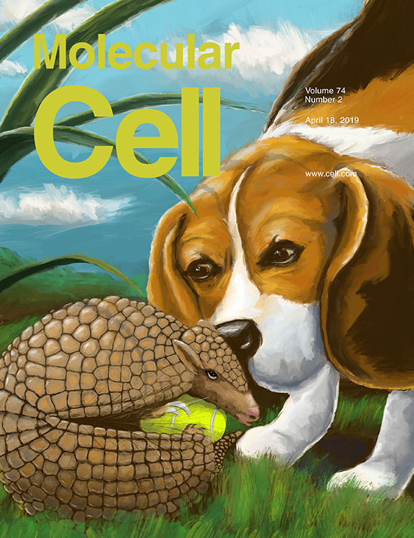
Bill Murray’s ominous premonition aside, animal metaphors have been invading my work. Here, a quality control phosphatase (beagle) surveys Protein Kinase C (armadillo). Alexandra Newton’s lab showed that if Protein Kinase C relaxes its compact conformation, its phosphate (tennis ball) will be plucked off and the kinase will be rapidly degraded. Important because more beagles and fewer armadillos correlates with decreased survival in pancreatic cancer.
And next, when the client says that you can make the cover whatever you want, but by the way, the first author really really likes cats, this is what happens. Josh Figueroa’s lab showed that ligand exchange in a cobalt complex proceeds via an associative mechanism. In other words, if the leaving ligand left before the new ligand joined (or if the orange cat poised to jump leaves before the black cat gets there to take its place), the whole thing becomes too unstable.

Alexa please explain machine learning
I took a project for the Amazon Alexa science blog, largely to try to impress the hubs (totally worked). But it was great fun and a nice way to hone my After Effects chops a little. There will be more of these.
Genomic mosaicism, or why our neurons are like snowflakes
Happy New Year! I realized that I haven’t been posting many updates here because I’ve moved more toward using Instagram for that (@oreillyscienceart). This year I resolve to restore some loyalty to my website, starting with these two images that I made to highlight a recent Nature paper from the Chun lab at the Sanford-Burnham Prebys Medical Discovery Institute. Genomic Mosaicism is a phrase used to describe the fact that neurons from the same brain don’t necessarily share the same DNA sequence. (I was delighted to use this as an excuse to create a mosaic image.) The authors of this paper reveal that a major contributor to this mosaicism is scrambling of DNA sequences through homologous recombination. They envisioned it as a gene bursting out to produce thousands of different variants of a single gene, resulting in an explosion of genetic diversity. By studying this phenomenon in the context of APP, a protein involved in Alzheimer’s, they find a correlation between the extent of scrambling and the onset of Alzheimer’s disease. They also make the enticing discovery that reverse transcriptase is responsible for the scrambling events. That’s right, the same reverse transcriptase that we already have approved drugs for. Another intriguing anecdote is that it is very rare for people being treated for HIV with reverse transcriptase inhibitors to get Alzheimer’s disease. Hopefully testing of these drugs in the context of Alzheimer’s disease will begin soon.
Somatic APP gene recombination in Alzheimer's disease and normal neurons.
Lee MH, Siddoway B, Kaeser GE, Segota I, Rivera R, Romanow WJ, Liu CS, Park C, Kennedy G, Long T, Chun J.
Nature. 2018 Nov;563(7733):639-645. doi: 10.1038/s41586-018-0718-6. Epub 2018 Nov 21.


On bashing genetic mailboxes
My Bauhaus-inspired cover came out in JACS last week. It’s about a protein chimera composed of a mutagenesis agent hitching a ride on an RNA polymerase to make mutations in specific genes. It reminded me of teenagers hanging out of the windows of moving cars, hitting mailboxes with baseball bats. (This metaphor probably dates me) In this case, the Shoulders Lab at MIT was clever enough to assign the teenagers (the mutagen) to a specific street. This image was made entirely on my iPad, which was a refreshing departure. Special thanks to George Howell Coffee in Newton, whose counter was the perfect height for drawing.

This cover design actually came up back in August when I was being interviewed by Carmen Drahl of Chemical and Engineering News at the ACS Boston meeting (seen here).

Obviously I didn’t say anything about the paper that hadn’t come out yet. She asked what my illustrations looked like, and I was explaining that the styles of my projects can vary dramatically. Even within this project, the other design we submitted for consideration for the cover was something much more typical of previous JACS covers (seen below). I was pretty surprised that they chose the wild card.

Hydra vulgaris is way better than it sounds
I had a nice surprise recently when this cover I made for Jacob T. Robinson's lab at Rice University came out. I didn't even know it had been chosen. It’s about immobilizing hydra (not the Marvel comics one) in a microfluidic device so its neurons can be poked and prodded. These little creatures are so cool. They stick to something at one end and then wave their little tentacles around, like a microscopic wacky wavy inflatable tube man. Or even better, like Dee from It's Always Sunny when she learns some new dance moves from one.

Taking on the challenge of scale from a different angle
Once again, I was faced with the task of trying to convey both the macroscopic and molecular in one image. The last time I talked about this (two posts ago), I used a combination of 2D and 3D. This time on the molecular level I needed to show structures of polymers, highlighting the halogen bonds (the cracks in the wall as well as the theme of the issue). On the macroscopic level I needed the metal balls grinding together two different polymers that would then crystallize into a co-polymer in the solid state (that's a thing!?). So, I decided to use shadows, the idea being that by imagining a light source very low and close to the crystal, I could exaggerate the shadow size and show the molecular details of the halogen-bonded polymers. Obviously I took a little artistic license with the shadow of the powder falling into an atom, but that's what journal covers are for.

Shortly after this project, I discovered Vincent Bal, who makes drawings from the shadows of everyday objects, incorporating the objects into the picture. I highly recommend checking these out unless you are an artist and you don't want to look at something that's going to make you want to quit forever, because they are that good:
https://www.instagram.com/vincent_bal/?hl=en
Is Zen doodling on deadline defeating the purpose?
This illustration was done for a paper about paneth cells de-differentiating into stem cells. A sort of paradigm in stem cell research until recently was Waddington’s concept of the ‘epigenetic landscape’, which depicts cells rolling down a hill as they differentiate from stem cells to their terminal cell types. It was long believed until recently that cells could not reverse this process, or roll back up the hill. In the paper this image depicts, the authors demonstrate that activation of the Notch1 pathway directly or via irradiation induces Notch-dependent acquisition of a stem cell-like transcriptome. Like Sisyphus pushing his rock up the hill, Notch is seen here pushing the Paneth cell back up the ‘epigenetic landscape’ to a de-differentiated state with stem cell features. Taken together these results suggest that Notch may play an important role in the Sisyphean task of intestinal epithelial cell regeneration, particularly in cases of inflammation or injury.

As my first attempt at Zen doodling I can tell you there was nothing Zen-like about it. But I learned a lot about making these and I would love to do more, with enough time. As an aside, the black and white version made great coloring pages for my kids. It occupied them for at least five minutes. No, I don't think they achieved "flow".
2D meets 3D
One of the great benefits of having moved back to Boston almost a year ago is that I've been able to reconnect with old grad school friends, one of whom hired me for this cover art project. It's about using mRNA sequencing to monitor yeast fermentation, reporting back when the system is low in some key component such as, in this case, lipids. The idea for this image was conceived of most pleasantly over sandwiches on a roof garden in Kendall Square, as opposed to my usual forehead-to-desk-alone-in-my-office method.

One challenge that comes up in the majority of my projects in the problem of scale. How do you represent objects that differ in scale by several orders of magnitude together in one image? Sometimes artists will use a magnifying glass, or even just an inset. Sometimes I will use extreme perspective. For this project I decided to try something new, and used 2D for the molecular and microscopic, while using 3D for the macroscale objects. It's meant as a sort of wink to the audience, as in, I know mRNA is not as big as a computer monitor but I put it in a different dimension so it's okay, see? Like a parallel universe! Will you allow it?
The pneumonia project
What are the odds that while working on a project creating an image of a bacterial cell wall, I would come down with pneumonia? (It was back in November and I'm fine now.) Which is how this henceforth became known as the pneumonia project. And it is why I had to complete it largely in bed, using my iPad to "paint" it. But I actually liked it much better than how it was shaping up in my fancy 3D modeling program, so, thanks pneumonia.

Which hiker are you?

I am definitely the one with the shoe untied and backpack unzipped. The hiking metaphor here is about varying levels of fitness for viral proteins accomplishing the task of protein folding. The fittest don't need any help from chaperones, while some are so unfit they get degraded before they even try folding. The idea for the hiking metaphor came from the first author of this paper, Angela Phillips, from Matthew Shoulders' lab at MIT. This was her original sketch for the concept:

In other news, we managed to have the O'Reilly Science Art holiday party of two this year. After a beer at the Cambridge Brewing Company, which I hadn't been to since I defended my thesis *mumblemumble* years ago, we saw Ladybird at the Kendall Square cinema. Because I am the boss. Or because the only showing of Star Wars The Last Jedi we could see started too late.
The Art of Basic Science #10
This painting came home from Kindergarten, and I was overcome with envy at this display of immune cells. Despite the fact that he had no idea that he was painting cells, I'd been out-science illustrated by my five year old, and so I thought this warranted a guest post by him again.

The Art of Basic Science #9
Otto Warburg was a Nobel Prize-winning German biochemist who championed the hypothesis, which we now know as the Warburg Hypothesis, that cancer is caused by cells switching from the respiration of oxygen to the fermentation of sugar. This was in 1924. This criteria has largely been relegated to a correlation, since with the advent of molecular biology we learned about mutations in DNA. It has been a controversial topic, and for what it's worth, there has been a 10-fold increase in articles related to the Warburg Effect over the past ten years.
In this study (see reference below image), the authors link yeast cell fermentation to the oncogene Ras. They not only correlate an influx of glucose with accumulation of fructose 1,6-bisphosphate and activation of Ras, they show that fructose 1,6-bisphophate triggers activation of Ras. This supports the Warburg Effect within the modern context of at least one way in which we understand cancer to work. The image here incorporates glucose, fructose-1,6-bisphosphate, proliferating cells, and Ras into a portrait of Otto Warburg. Only after the completion of this illustration did I realize that he studied chemistry under Emil Fischer, known among other things for drawing sugars in, that's right, Fischer projections. Had he studied under English chemist Sir Norman Haworth then it would have been apropos indeed. So it goes.

Fructose-1,6-bisphosphate couples glycolytic flux to activation of Ras
Ken Peeters, Frederik Van Leemputte, Baptiste Fischer, Beatriz M. Bonini, Hector Quezada, Maksym Tsytlonok, Dorien Haesen, Ward Vanthienen, Nuno Bernardes, Carmen Bravo Gonzalez-Blas, Veerle Janssens, Peter Tompa, Wim Versées, and Johan M. Thevelein
Nat Commun. 2017; 8: 922.
Published online 2017 Oct 13. doi: 10.1038/s41467-017-01019-z
It's a banner day!
This week a paper came out in Science Translational Medicine that describes the use of hematopoietic stem and progenitor cells to treat Friedrich's Ataxia. Once they differentiate into mature microglia (see in yellow), they can actually transfer proteins that are missing in Friedrich's Ataxia to the host's cells. They also differentiate into other cells in other parts of the body to deliver these rescue proteins as well. Pretty amazing stuff. I made this image for the Cherqui Lab at UCSD, the authors of the study, and you can see the image (for now anyway) as the second in the scrolling banner images on both the Science Translational Medicine and Science homepages:

Brighter, stronger, tunier - new flourophores from the Lavis Lab
The Lavis Lab found that adding the four-membered functional group known as azetidines to the classic rhodamine dyes makes the brighter and more photostable. Even cooler, they found that by functionalizing the azetidines they could fine-tune the properties of the dyes, and gave them a whirl in cells too.
The October installation of the Art of Basic Science is in the works. More soon!
